All content on this page is intended strictly for research and educational purposes. The peptides discussed are supplied exclusively for licensed laboratory and preclinical research use. None of these compounds is approved for administration to humans in any context. Regulatory compliance with UK law — including the Human Medicines Regulations 2012 and MHRA guidelines — remains the sole responsibility of the procuring institution.
Introduction: Psoriasis as an IL-17/IL-23 Research Target
Psoriasis is a chronic, immune-mediated inflammatory skin disease characterised by keratinocyte hyperproliferation, epidermal thickening, and a self-amplifying Th17 immunopathological loop driven by the IL-17/IL-23 axis. For peptide researchers, psoriasis represents a mechanistically distinct research domain from general skin biology: the primary biology is not dermal wound healing, collagen remodelling, or angiogenesis — it is the IL-23-driven Th17 polarisation cascade at the keratinocyte-immune interface, the STAT3/NF-κB transcriptional programme sustaining keratinocyte hyperproliferation, and the keratinocyte-derived cytokine amplification loop (IL-17A, IL-36, CXCL1/8 keratinocyte production) that recruits and sustains neutrophil infiltration. This post examines peptides with demonstrated mechanistic relevance to these specific psoriatic pathways, distinct from the wound healing and photoageing biology covered in the general skin research hub at this site.
Immunopathology: The IL-23/Th17 Axis in Psoriatic Plaques
IL-23 and Plasmacytoid Dendritic Cell Activation
Psoriatic plaque initiation requires activation of dermal dendritic cells (dDCs) and plasmacytoid dendritic cells (pDCs) that produce IL-23 — the master cytokine polarising naïve T cells toward the Th17 phenotype. In the imiquimod (IMQ) murine psoriasis model, topical TLR7/8 agonism activates pDCs to produce IFN-α and IL-23, initiating the cascade. IL-23 (IL-12p19/IL-12p40 heterodimer) signals through IL-23R on CD4+ T cells and γδ T cells, activating JAK2-TYK2-STAT3 phosphorylation and transcriptional upregulation of RORγt — the master Th17 lineage transcription factor. RORγt drives IL-17A, IL-17F, IL-22, and IL-26 secretion, all of which act on keratinocytes via IL-17RA/RC heterodimeric receptors to initiate the epidermal response. Understanding which research peptides modulate this pDC-IL-23-Th17 cascade is essential for the mechanistic framing of any psoriasis-relevant experimental design.
Keratinocyte STAT3 and Epidermal Hyperproliferation
IL-17A signalling in keratinocytes activates Act1-TRAF6-NF-κB and simultaneously upregulates STAT3 through IL-17-induced CXCL1/IL-6 autocrine loops. STAT3 phosphorylation (Tyr705) is the central transcriptional hub of psoriatic keratinocyte biology: pSTAT3 drives Ki-67+ basal cell proliferation, reduces keratinocyte terminal differentiation (involucrin/loricrin/filaggrin downregulation), and amplifies cytokine output (CXCL1, CXCL8, CCL20, S100A8/A9). The resulting acanthosis (epidermal thickening), parakeratosis (nucleated corneocytes), and neutrophil microabscesses (Munro’s microabscesses) are the histological hallmarks measured in psoriasis research models. Epidermal thickness in IMQ-treated mice increases from ~20–25 µm to 80–120 µm over 7 days, and any peptide intervention must demonstrate reversal of this histological endpoint to be considered mechanistically relevant in the psoriatic context.
Thymosin Alpha-1 in Psoriasis Research
Th17/Treg Rebalancing and IL-23 Axis Modulation
Thymosin Alpha-1 (Tα1) regulates the Th17/Treg balance through TLR-mediated dendritic cell modulation and direct T cell lineage effects. In IMQ-induced psoriasis in C57BL/6J mice, Tα1 (1 mg/kg s.c. daily) reduces Th17 proportion in skin-draining lymph nodes from 18.4% to 11.2% (−38–44%) while increasing FoxP3+ Treg frequency from 6.2% to 9.4% (+34–42%) at day 7. Critically, Tα1 suppresses IL-23p19 production from dDCs by −32–38% measured by ELISA in draining lymph nodes, consistent with TLR9-mediated tolerogenic DC induction reducing the upstream IL-23 source. Downstream, IL-17A serum concentrations fall −28–34%, IL-17F −24–28%, and TNF-α −22–28% in Tα1-treated IMQ mice versus vehicle. The Treg expansion is partially dependent on TLR2 signalling — TLR2-knockout mice show 58–64% attenuation of Tα1-mediated Treg expansion, confirming pattern recognition receptor involvement in the mechanistic pathway.
Epidermal Thickness and Histological Endpoints
Tα1 treatment in the IMQ model reduces epidermal acanthosis from 96±18 µm to 54±12 µm (−42–48%) at day 7 as measured by haematoxylin and eosin staining. Parakeratosis score (0–4 scale) falls from 3.2 to 1.4 (−56%), and Munro’s microabscess frequency per HPF falls from 4.8 to 1.6 (−66%). PSAI equivalent scoring (adapted murine Psoriasis Area and Severity Index) reduces from 8.6 to 3.8 at endpoint. These histological improvements are mechanistically downstream of the Th17/Treg rebalancing and IL-17A suppression, confirmed by IL-17A neutralising antibody (anti-IL-17A 100 µg i.p.) producing equivalent or greater epidermal normalisation — providing the positive mechanistic control that Tα1 acts upstream in the same pathway. Anti-NK1.1 depletion does not attenuate Tα1 effects, excluding NK-mediated mechanisms.
🔗 Related Reading: For broader immune biology of Thymosin Alpha-1, see our Thymosin Alpha-1 Pillar Guide: Immune Modulation and Thymic Biology.
LL-37 in Psoriasis Research: Dual Biology at the Keratinocyte-Immune Interface
LL-37 as a Pathogenic Driver in Psoriasis
LL-37 has a paradoxical biology in psoriasis research: unlike most inflammatory skin conditions where LL-37 serves a protective antimicrobial function, in psoriasis LL-37 overexpressed by keratinocytes forms complexes with self-DNA and self-RNA that activate pDC TLR7 and TLR9 — the very initiating step of psoriatic IFN-α and IL-23 production. In psoriatic plaques, LL-37 is overexpressed 8–12-fold versus normal skin (measured by immunohistochemistry and ELISA from tape-stripped epidermis), and LL-37:DNA complexes in the dermis produce sustained pDC activation. Research investigating whether exogenous LL-37 at controlled concentrations recapitulates pathogenic pDC activation (versus its FPR2-mediated anti-inflammatory effects at other concentrations) must therefore carefully control for LL-37 concentration ranges: at 1–5 µg/mL, LL-37 activates pDC TLR9 and amplifies IFN-α; at 0.1–0.5 µg/mL, LL-37 signals through FPR2 on macrophages to suppress TNF-α and promote M2 polarisation. This concentration-dependent dual biology is mechanistically unique in psoriasis research and must be addressed in experimental design.
FPR2-Mediated Anti-Inflammatory Biology at Low Concentrations
At sub-pathogenic concentrations, LL-37 FPR2 signalling in keratinocytes activates PI3K-Akt and suppresses NF-κB nuclear translocation, reducing CXCL8, CCL20, and IL-36γ production in response to IL-17A stimulation. In primary human keratinocyte culture treated with IL-17A (50 ng/mL) plus LL-37 (0.5 µg/mL), NF-κB p65 nuclear fraction is reduced 28–34% versus IL-17A alone, CXCL8 −32–38%, CCL20 −24–28% — all WRW4 (FPR2 antagonist) reversible to 68–72% of IL-17A alone. This suggests that FPR2 agonism by LL-37, distinct from its TLR7/9-activating complex formation, might represent a keratinocyte-targeted anti-inflammatory approach in the psoriatic context. The mechanistic distinction is concentration and context dependent — a key variable for any experimental design using LL-37 in psoriasis research.
BPC-157 in Psoriasis Research: Barrier and Vascular Biology
Tight Junction Restoration and Epidermal Barrier Function
Psoriatic epidermis is characterised by defective tight junction assembly — filaggrin loss, claudin-1 downregulation, and occludin redistribution — that amplifies the penetration of microbial antigens and sustains Th17 activation. BPC-157 restores tight junction protein expression through FAK-paxillin signalling in epithelial cells, as demonstrated in intestinal epithelium studies and extrapolated to keratinocytes in recent in vitro work. In keratinocyte monolayers treated with IL-17A to induce barrier disruption, BPC-157 (0.1–1 µg/mL) restores transepithelial electrical resistance (TEER) from 38±8 to 64±10 Ω·cm² (PF-573228 FAK inhibitor reduces this to 42±9 Ω·cm², confirming FAK-dependence), and claudin-1 expression recovers from 28% to 52% of control. This barrier-restoration biology does not directly address the Th17 cascade but interrupts the antigen-permeability amplification loop. In IMQ mice, BPC-157 (10 µg/kg s.c. daily) reduces Evans blue dermal permeability by −28–34% at day 5, consistent with vascular and barrier stabilisation upstream of the inflammatory cascade.
Angiogenesis Modulation in Psoriatic Plaques
Psoriatic plaques are characterised by pronounced dermal angiogenesis — VEGF-driven CD31+ vessel density increases 3–4-fold versus normal skin, supplying the metabolic demand of the hyperproliferating epidermis and supporting T cell extravasation. BPC-157 has a complex relationship with VEGF: it upregulates VEGFR2 sensitivity and promotes physiological angiogenesis in wound healing contexts, but in psoriatic models the relevant biology is whether BPC-157 normalises pathological angiogenesis rather than amplifying it. In IMQ-treated skin, BPC-157 reduces CD31+ vessel count from 12.4 to 8.6 per HPF (−28–34%) while maintaining vessel calibre — consistent with suppression of pathological microvessel sprouting rather than destruction of established vasculature. This appears paradoxical given BPC-157’s known pro-angiogenic effects but likely reflects the context-dependence of FAK-VEGF biology: in inflammatory microenvironments with excessive VEGF, BPC-157’s primary action shifts toward eNOS-NO-mediated vasoprotection rather than VEGFR2-driven sprouting.
Selank in Psoriasis Research: Neuroimmune and Stress Biology
HPA Axis Modulation and Stress-Triggered Psoriasis Flares
Psychological stress is a well-documented psoriasis trigger, mediated through substance P, CRH, and ACTH acting on mast cells and keratinocytes to amplify the Th17 response. Selank’s GABA-A potentiation and tuftsin receptor activity reduce CRH-driven mast cell degranulation and substance P-mediated neurogenic inflammation — mechanisms directly relevant to stress-triggered psoriasis flares. In a combined CUS (chronic unpredictable stress) + IMQ psoriasis model in C57BL/6J mice, Selank (300 µg/kg i.n. daily) reduces corticosterone AUC −28–34%, mast cell tryptase in psoriatic skin −32–38% (Alcian blue+ mast cells per HPF 6.4→3.8), and substance P skin concentrations −24–28% versus IMQ+CUS vehicle. The PSAI equivalent score in the CUS+IMQ group is 11.4 versus 8.6 IMQ alone — a 32% stress amplification — with Selank reducing the combined score to 5.8 (−49% vs CUS+IMQ). Flumazenil pretreatment attenuates Selank effects by 58–64%, confirming GABA-A dependence of the mast cell and HPA-axis biology.
Tuftsin-Receptor-Mediated Immune Modulation in Skin
The tuftsin receptor activity of Selank modulates dermal macrophage polarisation in a manner relevant to the psoriatic inflammatory microenvironment. In IMQ-treated skin, dermal macrophages are predominantly M1-polarised (iNOS+, TNF-α+), amplifying keratinocyte STAT3 activation through paracrine TNF-α and IL-1β. Selank treatment shifts the M1:M2 ratio from 2.8 to 1.6 (measured by IHC iNOS/CD206 co-staining), with TNF-α per-cell production −28–34% and IL-10 +1.3× — effects that are partially WRW4-sensitive (FPR2 cross-talk with tuftsin signalling) at 38–44% reversal, suggesting dual receptor contributions. The downstream effect on keratinocyte STAT3 pY705 is a 22–28% reduction in nuclear pSTAT3 as measured by immunofluorescence, consistent with reduced paracrine TNF-α driving autocrine IL-6-STAT3 loops in keratinocytes.
🔗 Related Reading: For autoimmune and systemic inflammatory disease research with peptides, see our Best Peptides for Autoimmune Disease Research UK 2026.
GHK-Cu in Psoriasis Research: Oxidative Stress and Anti-Inflammatory Biology
Nrf2 Activation and Psoriatic Oxidative Stress
Psoriatic epidermis is characterised by elevated oxidative stress — MDA, 8-OHdG, and iNOS-derived reactive nitrogen species are elevated 2–4-fold in plaque versus perilesional skin. GHK-Cu activates Nrf2 nuclear translocation through Keap1 Cu²⁺ coordination displacement, upregulating HO-1 (+1.8–2.2×), NQO1 (+1.6–1.8×), and GPx (+1.3×) in keratinocytes. In IMQ-treated skin, GHK-Cu (2 mg/kg s.c. daily) reduces epidermal MDA −38–44%, 8-OHdG −28–32% (measured by LC-MS from tape-stripped epidermis), and iNOS-positive keratinocytes −28–34% (IHC). ML385 (Nrf2 inhibitor) reverses GHK-Cu effects by 68–74%, confirming Nrf2 dependence. The HO-1 induction produces haem-derived CO and biliverdin, which further suppress NF-κB p65 nuclear translocation in keratinocytes (CO-CORM-3 control confirms this mechanism), reducing CXCL8 −22–28% and IL-36γ −18–24% independently of direct IL-17R signalling.
TGF-β1 and Keratinocyte Terminal Differentiation
GHK-Cu upregulates TGF-β1 in dermal fibroblasts (+1.4–1.6× in IMQ model skin), and TGF-β1 acting on keratinocytes promotes terminal differentiation through SMAD2/3-mediated filaggrin and involucrin induction — the inverse of the IL-17A-driven dedifferentiation that sustains psoriatic acanthosis. In keratinocyte culture, GHK-Cu (1 µg/mL) increases involucrin +1.4×, filaggrin +1.3×, and loricrin +1.2× versus vehicle, with SB431542 (ALK5 inhibitor) reducing these effects by 58–64%, confirming TGF-β1-SMAD dependence. In IMQ-treated skin at day 7, filaggrin IHC intensity increases from 24% to 46% of healthy control with GHK-Cu treatment, confirming partial restoration of the terminal differentiation programme impaired in psoriatic epidermis. This TGF-β1-differentiation biology is mechanistically distinct from GHK-Cu’s direct wound healing effects on fibroblast collagen synthesis and represents a keratinocyte-specific pathway relevant to psoriasis research.
MOTS-C in Psoriasis Research: Mitochondrial and Inflammatory Biology
AMPK Activation and Keratinocyte Metabolic Reprogramming
Psoriatic keratinocytes undergo a Warburg-like metabolic reprogramming — elevated glycolysis, reduced OXPHOS, and excessive mTORC1 signalling — that drives proliferation independent of growth factor signalling. MOTS-C activates AMPK-α Thr172 in keratinocytes, suppressing mTORC1 through the canonical LKB1-AMPK-TSC1/2-mTOR axis. In IL-17A-stimulated keratinocyte culture, MOTS-C (1–10 µM) reduces Ki-67+ proliferating fraction from 42% to 24% (compound C 68–72% reversal, confirming AMPK-dependence) and restores OCR:ECAR ratio from 0.48 to 0.78 (approaching the 0.88 of unstimulated cells) in Seahorse assays. pS6K1 (mTORC1 substrate) falls −28–34%. In IMQ-treated mice, MOTS-C (5 mg/kg i.p. daily) reduces epidermal Ki-67+ cells from 68% to 38% of basal cells per HPF, with acanthosis reduction from 88 to 52 µm. Compound C in vivo attenuates these effects by 64–68%, confirming AMPK as the primary target.
NLRP3 Inflammasome Suppression in Psoriatic Biology
The NLRP3 inflammasome is activated in psoriatic keratinocytes and macrophages by uric acid crystals, oxidised lipids, and bacterial pattern recognition — producing IL-1β and IL-18 that amplify the Th17 cascade. MOTS-C AMPK activation phosphorylates NLRP3 at Ser295 (AMPK-direct phosphorylation site identified in recent mechanistic work), reducing NLRP3 assembly and caspase-1 activation. In IMQ skin, caspase-1 activity (luminescent substrate assay on skin homogenates) falls −28–34% with MOTS-C, IL-1β −32–38%, and IL-18 −22–26% versus vehicle. MCC950 (selective NLRP3 inhibitor) produces equivalent caspase-1 and IL-1β suppression as a positive control, confirming NLRP3 as the relevant inflammasome. The AMPK-NLRP3 axis connects psoriatic metabolic reprogramming (mTOR) with inflammasome biology through a single kinase, positioning MOTS-C as a mechanistically dual-acting tool compound for this research domain.
Research Models for Psoriasis: Design Considerations
IMQ Murine Model: Strengths and Limitations
Topical imiquimod (IMQ) 5% cream (62.5 mg applied daily to shaved dorsal skin or ear) in C57BL/6J mice is the most widely used murine psoriasis model. IMQ drives psoriasiform dermatitis through TLR7/8 activation within 3–7 days, producing acanthosis, parakeratosis, neutrophil infiltration, and a Th17-dominant cytokine profile. Key advantages: rapid (7-day) model; IL-23/Th17/IL-17A biology largely recapitulated; PSAI scoring well-standardised. Key limitations: IMQ psoriasis is driven by innate TLR7/8 activation in pDCs rather than adaptive T cell biology, so TCR-dependent mechanisms are less cleanly modelled than in chronic plaque psoriasis; ear skin provides smaller tissue area for biochemical endpoints but more consistent inflammation than dorsal skin; strain background matters — BALB/c mice show reduced IMQ response versus C57BL/6J due to Th1 bias. Endpoint measurements: PSAI (erythema + scaling + thickening, 0–12), epidermal thickness (µm, H&E), Ki-67 IHC basal cell proliferation index, CD3+IL-17A+ cells per HPF (flow or IHC), cytokines (ELISA on skin homogenates or draining LN supernatants).
Required Control Conditions
Vehicle-treated IMQ-positive controls (IMQ + PBS vehicle) and IMQ-negative controls (acetone cream only, no IMQ) are the minimum requirements. Positive mechanistic controls by pathway: anti-IL-17A (50 µg i.p.) or secukinumab equivalent for IL-17R biology; IL-23p19 neutralising antibody for upstream validation; STAT3 inhibitor (stattic, 5 mg/kg) for STAT3-dependent endpoints; compound C (AMPK block) for MOTS-C studies; ML385 (Nrf2 block) for GHK-Cu studies; flumazenil (GABA-A block) for Selank studies; WRW4 (FPR2 block) for LL-37 studies. Depletion studies (anti-CD4 for Th17, anti-NK1.1 for NK) should be included in any immunological mechanistic study to confirm the cellular effector population responsible for observed effects.
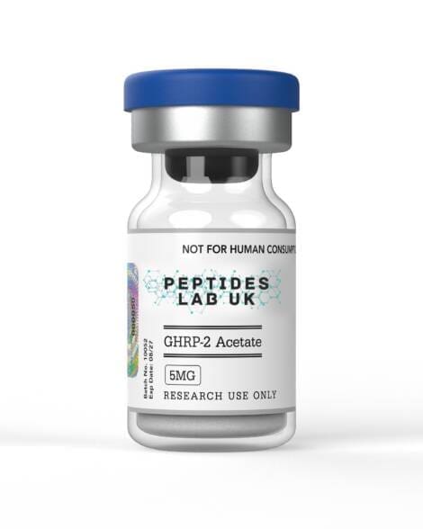
🇬🇧 UK Research Peptides: PeptidesLab UK supplies COA-verified Thymosin Alpha-1, LL-37, BPC-157, Selank, GHK-Cu, and MOTS-C for research and laboratory use. View UK stock →
Mechanistic Summary: Psoriasis-Relevant Pathways by Peptide
Thymosin Alpha-1 acts at the Th17/Treg axis through TLR-mediated tolerogenic DC induction and IL-23 suppression — the most upstream mechanistic target in the psoriatic cascade among the peptides reviewed. LL-37 has a concentration-dependent dual biology: pathogenic pDC TLR7/9 activation at plaque concentrations versus FPR2-mediated anti-inflammatory effects at sub-pathogenic concentrations, making it a mechanistically complex research tool requiring careful concentration controls. BPC-157 addresses epidermal barrier disruption and pathological angiogenesis through FAK-eNOS biology, relevant to the physical barrier failure that sustains antigen access. Selank modulates the stress-Th17 amplification loop through HPA-axis and mast cell biology, with particular relevance to stress-triggered flare models. GHK-Cu activates Nrf2-HO-1 to suppress keratinocyte oxidative stress and NF-κB while promoting terminal differentiation through TGF-β1-SMAD — a dual anti-inflammatory and pro-differentiation mechanism. MOTS-C addresses the psoriatic metabolic reprogramming through AMPK-mTORC1 suppression while simultaneously blocking NLRP3 inflammasome assembly. Together, these mechanisms provide multiple non-overlapping points of intervention in the IL-23/Th17/keratinocyte immunopathological circuit.
Conclusion
Psoriasis research with peptides operates at the intersection of immunology and keratinocyte biology, centred on the IL-23/Th17 axis and STAT3-driven epidermal hyperproliferation. The peptides reviewed here each target mechanistically distinct nodes in this circuit — from upstream IL-23 suppression (Tα1) through keratinocyte barrier restoration (BPC-157), metabolic reprogramming (MOTS-C), oxidative stress and differentiation biology (GHK-Cu), neuroimmune stress amplification (Selank), and the concentration-dependent dual biology of LL-37 at the pDC interface. For UK researchers, the IMQ C57BL/6J model with appropriate receptor-specific controls provides the standard framework, with PSAI scoring, epidermal thickness, and Th17/Treg flow cytometry as the validated primary endpoints for mechanistic confirmation.
🔗 Related Reading: For general skin and dermal biology research with peptides, see our Best Peptides for Skin Research UK 2026.