Important regulatory notice. No peptide sold as a research-use-only reference compound is licensed by the MHRA as a skin-ageing or anti-ageing medicine in the United Kingdom. This page is a literature-context overview of compound families discussed in published skin-ageing research. It is not personal-use guidance. Peptides Lab UK supplies research-use-only laboratory reference compounds. Products are not for human or veterinary use.
Quick research summary. The published skin-ageing literature has explored several peptide and peptide-related compound families in cell-culture and small-animal model contexts, covering photoageing biology, dermal matrix remodelling, fibroblast senescence, and pigmentation biology. The compounds discussed appear in that research record. Cosmetic peptide products containing some of these families are sold separately as regulated cosmetics where they make only cosmetic claims.
Skin-ageing biology context
Skin-ageing literature describes both intrinsic chronological ageing (telomere attrition, collagen and elastin reduction, fibroblast senescence) and extrinsic ageing (UV-induced photoageing, oxidative stress, glycation, environmental damage). The dermal matrix biology, fibroblast biology and melanocyte biology each have their own substantial peer-reviewed literature.
Compound families that appear in the published skin-ageing research record
Cell-culture and animal-model studies have discussed several peptide families. Copper peptides such as GHK-Cu appear in collagen-biochemistry and dermal-fibroblast research. Matrikine peptides (Matrixyl and similar) appear in dermal matrix research. Antioxidant and anti-glycation peptides appear in oxidative-biology research. Pigmentation-biology peptide research includes melanocortin-pathway compounds.
The cosmetic vs research-use distinction
Topical cosmetic products containing some of the peptide families discussed above are sold under cosmetics regulation and make cosmetic claims only. They are not the same product category as research-use-only laboratory reference compounds. Peptides Lab UK supplies research-use-only reference compounds, not cosmetic products.
UK regulatory position
No research-use-only peptide on this site is a licensed treatment for skin ageing. Cosmetic products with cosmetic claims are regulated under cosmetics law. The MHRA position on therapeutic claims about unregulated peptides applies to any seller making medicinal claims about skin-ageing peptides outside the cosmetics framework.
For laboratory researchers
Skin-biology researchers may use peptide reference compounds for in-vitro and ex-vivo skin-model studies. Quality requirements are batch-specific certificate of analysis, third-party HPLC purity data, mass-spectrometry identity confirmation, and clear research-use-only labelling.
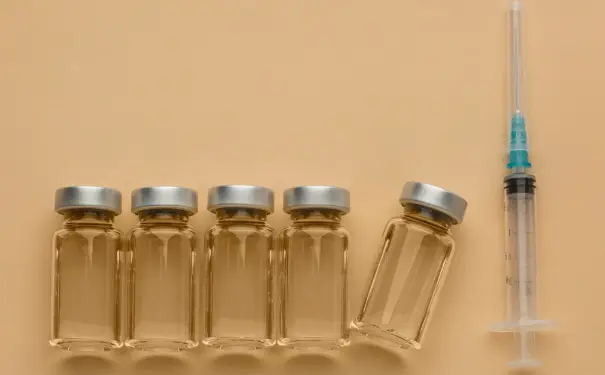
Research use only. Peptides Lab UK supplies research-use-only laboratory reference compounds with batch-specific certificates of analysis. Products are not for human or veterinary use. Cosmetic peptide products are sold separately under cosmetics regulation by other retailers.