Quick Answer Box: Both are neuropeptides produced in the hypothalamus, but oxytocin primarily drives trust, maternal bonding, and empathy, while vasopressin governs territorial behavior, pair-bond fidelity, and long-term mate guarding — especially in males.
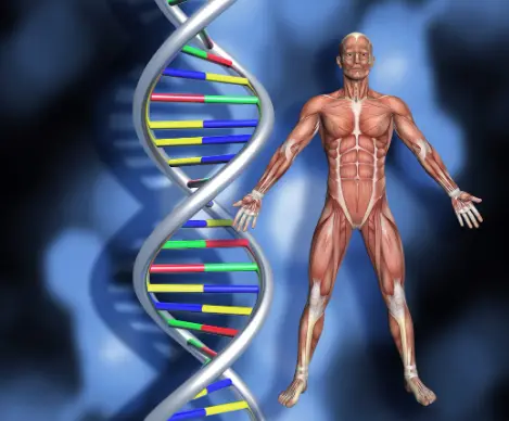
When researchers began exploring the neuroscience of human connection, they kept arriving at two small but profoundly powerful molecules: oxytocin and vasopressin. Both are neuropeptides synthesized in the hypothalamus, and both have become central to scientific discussions about why humans form deep emotional bonds, how trust is established between strangers, and what drives long-term commitment in romantic relationships. Yet despite their shared origin and structural similarity — differing by only two amino acids — the two molecules operate through distinct receptor systems, influence different brain regions, and produce fascinatingly different behavioral outcomes.
Oxytocin has earned significant media attention over the past two decades, often described in popular science as the ‘love hormone’ or ‘bonding molecule.’ This characterization, while somewhat reductive, reflects genuine findings from clinical research and animal studies. When oxytocin is released during childbirth, breastfeeding, or physical touch, it triggers a cascade of neurochemical events that promote closeness and reduce social anxiety. Understanding how this neuropeptide works at a molecular level — and how it compares to its structural cousin vasopressin — is essential for anyone interested in the biology of human relationships.
Vasopressin, historically studied for its role in water regulation and blood pressure, is equally important in shaping social behavior, particularly in males. It influences aggression in competitive contexts, supports fidelity in pair-bonded species, and activates reward pathways associated with long-term mate preference. The interplay between these two systems is now regarded as one of the most important frontiers in social neuroscience. This article examines their biological origins, receptor systems, behavioral effects, and research findings, drawing on studies in rodents, primates, and humans to build a comprehensive picture of how they differ in the context of social bonding.
Table of Contents
The Biological Origins of Oxytocin and Vasopressin
Shared Synthesis, Divergent Functions
Both oxytocin and vasopressin are nonapeptides — composed of nine amino acids — and share seven of those nine residues. This structural overlap explains why they were frequently confused in early research and why each can weakly bind to the other’s receptor at high concentrations. Both are produced by magnocellular neurons in the paraventricular nucleus (PVN) and supraoptic nucleus (SON) of the hypothalamus, and both travel to the posterior pituitary gland for storage before being released into systemic circulation in response to specific physiological triggers.
Oxytocin release is stimulated by uterine contractions during labor, nipple stimulation during breastfeeding, and various forms of social touch including hugging and physical contact between bonded individuals. Once in the bloodstream, it acts as a peripheral hormone affecting the uterus, mammary glands, and cardiovascular system. Crucially, oxytocin is also released centrally — directly into brain regions including the amygdala, hippocampus, nucleus accumbens, and prefrontal cortex — where it shapes mood, social perception, and the motivation to connect with others.
What Triggers Vasopressin Release
Vasopressin responds to a different set of physiological signals. Increased blood osmolality, low blood volume, and physical stress are the primary stimuli for its release. In the brain, it operates through a network of regions that overlap with — but are distinct from — the oxytocin system, including the lateral septum, ventral pallidum, and bed nucleus of the stria terminalis. These regions are deeply implicated in social memory and reward processing, which helps explain vasopressin’s central role in long-term bonding behavior and partner recognition.
The evolutionary conservation of both peptides across virtually all vertebrate species underscores their biological importance. Structural homologs of oxytocin and vasopressin have been identified in species ranging from roundworms to primates, suggesting these neuropeptide systems for regulating social and reproductive behavior have existed for hundreds of millions of years — making them among the most ancient and conserved biological mechanisms for governing social life.
Receptor Systems, Brain Distribution, and Behavioral Differences
How Oxytocin Receptors Shape Social Reward
The behavioral differences between oxytocin and vasopressin arise largely from where and how their receptors are distributed across the brain. The oxytocin receptor (OXTR) is found in high concentrations in the nucleus accumbens, amygdala, and hypothalamus. Its presence in the nucleus accumbens — the brain’s primary reward hub — is particularly significant because it links social interaction with dopamine-driven pleasure, essentially making positive social experiences feel rewarding and worth repeating. This oxytocin-dopamine connection is considered a cornerstone mechanism by which social bonding becomes motivationally compelling.
The distribution of OXTR in the amygdala is equally important. Research consistently shows that oxytocin reduces amygdala reactivity to threatening social stimuli, which explains why it is associated with lower social anxiety and greater openness in social situations. Early life stress has been shown in animal models to alter OXTR expression, potentially programming lifelong differences in social behavior and attachment capacity — suggesting that the receptor landscape is shaped not just by genetics but by lived experience.
Vasopressin V1a Receptor Distribution and Monogamy
Vasopressin acts through three known receptor subtypes: V1a, V1b, and V2. The V1a receptor is most relevant to social behavior and is densely expressed in the lateral septum and ventral pallidum. Research on prairie voles and meadow voles — two closely related rodent species with dramatically different mating systems — demonstrated that differences in V1a receptor density in the ventral pallidum largely account for why prairie voles form lifelong monogamous pair bonds while meadow voles do not. This groundbreaking finding showed that receptor distribution, not the peptide itself, determines bonding behavior.
Genetic polymorphisms in the AVPR1A gene (encoding the V1a receptor) have been linked in human studies to differences in marital quality, partner bonding, and brain activation in response to social cues. A study published in PNAS found that men carrying a particular AVPR1A allele variant scored lower on measures of pair bonding and reported lower marital quality — as did their partners. These findings point to vasopressin receptor genetics as a meaningful, if partial, contributor to individual variation in human relationship depth.
Oxytocin’s Role in Maternal Bonding and Infant Attachment
Animal Models and the Maternal Brain
No area of oxytocin research has produced more consistent and compelling findings than its role in mother-infant bonding. In rodent models, blocking oxytocin receptors in the PVN immediately after delivery disrupts the formation of maternal behavior — mothers fail to build nests, retrieve pups, or adopt nursing postures. Conversely, infusing oxytocin into the brain of virgin female rats can induce maternal behavior without pregnancy or delivery. These results establish oxytocin as a critical molecular switch for activating the caregiving system in the mammalian brain.
In human research, plasma levels of oxytocin during the first weeks postpartum correlate with the frequency of maternal affectionate behaviors including vocalization, physical contact, and attentive monitoring of the infant. Mothers with higher oxytocin concentrations show greater activation in brain regions associated with reward and emotional salience when viewing images of their own child compared to unfamiliar infants. This suggests that oxytocin does not simply cause attachment — it amplifies the salience and reward value of a specific individual to the caregiver’s brain.
Oxytocin Released During Physical Touch and Romantic Bonding
The role of oxytocin in romantic pair bonding has been extensively studied. Research has found that couples who engage in physical touch — including hugging, hand-holding, and close proximity — show measurable increases in oxytocin levels. Studies using intranasal synthetic oxytocin have shown that male participants in committed relationships rated their own partner as more attractive and maintained greater physical distance from unfamiliar attractive women compared to those who received placebo. This partner-fidelity effect of oxytocin — functionally similar to what vasopressin achieves in prairie voles — suggests overlapping bonding mechanisms across the two peptide systems.
This fidelity effect is context-dependent: single men showed no avoidance response to unfamiliar women after oxytocin administration, indicating that the peptide interacts with existing relationship status rather than simply increasing attraction indiscriminately. This context-dependence is a recurring theme in oxytocin research and complicates popular narratives about it as a universally prosocial molecule. The research picture is more nuanced: oxytocin is better understood as a social salience amplifier — making whatever is most socially relevant in a given moment feel more vivid, rewarding, or significant.
How Oxytocin Affects Trust, Empathy, and Social Anxiety
Research Evidence on Oxytocin and Interpersonal Trust
One of the most cited findings in the oxytocin literature comes from a 2005 study published in Nature by Kosfeld and colleagues, which used an economic trust game to show that participants who received intranasal oxytocin were significantly more willing to entrust money to strangers than those receiving placebo. This study generated enormous interest in oxytocin as a potential modulator of generalized social trust. Subsequent research extended these findings into cooperation, negotiation, and social decision-making — areas where oxytocin consistently appears to lower the perceived social risk of trusting an unfamiliar person.
Subsequent studies added important nuance, however. Oxytocin appears to enhance trust selectively — toward in-group members more than out-group strangers. Research by De Dreu and colleagues demonstrated that oxytocin increased cooperation and favoritism toward one’s own group while simultaneously increasing defensive aggression toward perceived out-group competitors. This in-group favoritism effect suggests that oxytocin does not uniformly promote positive social behavior but instead functions to strengthen the social boundaries of existing groups — a finding with significant implications for understanding intergroup conflict.
Oxytocin and Social Anxiety: What the Neuroscience Reveals
At the neural level, oxytocin reduces amygdala reactivity to threatening social stimuli — a well-replicated finding from functional neuroimaging studies. Intranasal oxytocin attenuates blood-oxygen-level-dependent responses in the amygdala when participants view fearful or angry faces. Because amygdala hyperactivity is associated with social anxiety disorder and autism spectrum conditions, this finding has generated considerable interest in oxytocin as a potential therapeutic target. Clinical trials to date have produced mixed results, but the mechanistic evidence for an anxiolytic effect of oxytocin in social contexts remains strong.
The relationship between oxytocin and empathy is also well-established in laboratory settings. Research participants who received intranasal oxytocin showed improved performance on the ‘Reading the Mind in the Eyes’ test, a validated measure of the ability to infer others’ emotional states from facial cues. This enhancement in social perception may underlie broader effects on trust and prosocial behavior — if oxytocin makes it easier to accurately read and respond to others’ emotions, it naturally makes social interactions feel more rewarding and socially safer. The connection to oxytocin and stress response is also relevant here: oxytocin has been shown in multiple studies to attenuate cortisol release in stressful social situations, effectively reducing the physiological cost of social engagement.
Vasopressin and Pair-Bond Formation: Evidence from Animal Research
The Prairie Vole Model and Monogamy
The prairie vole (Microtus ochrogaster) has become one of the most important animal models in social neuroscience because of what it reveals about vasopressin and pair bonding. Prairie voles are among the few mammalian species that form lifelong monogamous pair bonds. After mating, males become strongly attached to their female partner, demonstrate partner preference in choice tests, and engage in mate guarding — behaviors that are largely absent in the closely related meadow vole (Microtus pennsylvanicus), which is polygamous.
Researchers traced this behavioral difference to V1a receptor expression in the ventral pallidum, a region dense with dopamine receptors forming part of the brain’s reward circuitry. When vasopressin is released during mating in the prairie vole, it binds to V1a receptors in the ventral pallidum, triggering dopamine release that creates a rewarding association specifically with the partner’s scent and presence. In meadow voles, V1a receptors are sparse in this region, so the same vasopressin release fails to produce the same partner-specific reward signal. Pair bonding, in this model, is essentially the conversion of a social encounter into a long-term reward memory.
Vasopressin and Social Memory in Human Bonding
What makes these vole findings compelling for human research is that the V1a receptor gene (AVPR1A) contains a repetitive microsatellite region in its promoter sequence that influences receptor expression levels. Prairie voles carry a longer version of this microsatellite, correlating with higher receptor density in the ventral pallidum. Humans carry variable-length versions of a similar microsatellite, and research has found associations between specific AVPR1A allele variants and lower pair-bond quality in men. Vasopressin’s role in social memory — the ability to remember and preferentially respond to familiar social partners — is also central to how long-term bonds are maintained once formed.
These findings do not imply genetic determinism. Environment, developmental history, and other biological systems all contribute to relationship quality. But they indicate that vasopressin signaling is a meaningful contributor to the neural architecture underlying human pair bonding, and that individual differences in this system may partly explain why some individuals form deep, enduring bonds while others struggle to maintain long-term attachment. The research suggests that vasopressin and social memory are functionally inseparable in the context of sustained romantic partnership.
Vasopressin’s Role in Male Social Behavior, Aggression, and Protection

Competitive Behavior and Mate Guarding
While oxytocin research has focused largely on affiliative and caregiving behaviors, vasopressin research has more frequently highlighted its role in competitive, territorial, and protective social behaviors — particularly in males. In animal models, vasopressin infused into the lateral septum or anterior hypothalamus increases aggressive responses, while blocking V1a receptors in these areas reduces aggression. This has led some researchers to conceptualize vasopressin as part of a broader system for coordinating social threat responses and defending valued social relationships.
The link between vasopressin and aggression is not straightforward, however. In many contexts, vasopressin facilitates mate guarding and partner-protective behavior rather than generalized hostility. A male prairie vole that has formed a pair bond shows heightened aggression toward unfamiliar males — a behavior that depends on vasopressin signaling in the lateral septum. In this context, what appears to be aggression is more accurately understood as a protective social function: defending an existing bond rather than expressing hostility toward the world at large.
Sex Differences in Vasopressin and Oxytocin Systems
In humans, research shows that men respond to social threat cues with increased vasopressin release, and this rise correlates with heightened attention to competitive social information. Functional neuroimaging studies have shown that intranasal vasopressin modulates amygdala and cingulate cortex activity in response to social signals in ways that differ from oxytocin, and these effects tend to be more pronounced in males. Sexual dimorphism in the vasopressin system is well-established: male brains typically show higher V1a receptor density in regions associated with mate guarding and competitive behavior, while female brains show greater OXTR sensitivity.
Some researchers propose that this represents an evolutionary division of labor: oxytocin predominantly supports mother-infant and female-female social bonding essential for cooperative childcare in ancestral environments, while vasopressin supports the mate-guarding and coalition-building behaviors more critical for male reproductive success. Both systems are present in both sexes, but their relative weighting differs, likely shaped by the interplay between sex hormones and neuropeptide receptor expression across development. Estrogen upregulates OXTR expression, while testosterone amplifies vasopressin’s social behavioral effects — meaning the balance between systems shifts across the lifespan.
How the Oxytocin and Vasopressin Systems Interact
Cooperative and Opposing Effects in the Brain
Oxytocin and vasopressin do not operate in isolation. Their receptor systems overlap in many brain regions, and the two peptides frequently modulate each other’s effects. In some brain areas, they appear to have opposing actions — oxytocin dampens anxiety and promotes social approach, while vasopressin in the same region promotes vigilance and social threat assessment. Research has proposed a contextual model in which oxytocin predominates under conditions of perceived safety, facilitating trust and affiliation, while vasopressin and its V1a receptor dominate under conditions of anxiety or threat, increasing defensiveness and social selectivity.
In other contexts, the two peptides work cooperatively, both contributing to the formation and maintenance of social bonds through complementary mechanisms. Research on prairie voles has shown that both peptides contribute to partner preference formation, but through distinct neural pathways. Oxytocin acting in the nucleus accumbens and vasopressin acting in the ventral pallidum both trigger dopamine release that reinforces partner-specific social reward. Blocking either system alone partially reduces pair bonding; blocking both together more completely eliminates it. This redundant architecture may reflect the evolutionary importance of reliable bonding mechanisms.
Epigenetic Tuning of Neuropeptide Receptors
Adding to this complexity, both the oxytocin receptor and the V1a vasopressin receptor are subject to epigenetic modification — meaning their expression can be altered by early life experiences without any change to the underlying DNA sequence. Animal studies have shown that early life stress, maternal separation, or neonatal neuropeptide exposure can produce lasting changes in OXTR and AVPR1A expression that shape social behavior in adulthood. This epigenetic tunability means the bonding capacity of an individual is not fixed at birth but is shaped by developmental experience — a finding with broad implications for understanding how early trauma or nurturing environments leave lasting marks on the biology of social attachment.
Oxytocin in Friendship, Cooperation, and Cross-Species Bonding
Beyond Romantic Love: Oxytocin in Everyday Social Connection
Oxytocin’s influence on social bonding extends well beyond romantic partnerships and mother-infant relationships. Research has demonstrated that oxytocin levels rise during positive social interactions between friends, during cooperative play in children, and during synchronized activities such as group singing, team rowing, or shared rhythmic movement. This broader role suggests that oxytocin is not a specialized bonding hormone for intimate relationships but a more general facilitator of positive social engagement across diverse relationship types. The neuropeptide social bonding brain research literature now consistently shows oxytocin rises in prosocial, cooperative, and mutually rewarding social contexts regardless of their romantic or familial nature.
Research on human-dog interaction has attracted considerable attention in this context. A study published in Science found that mutual gaze between humans and their pet dogs triggered measurable increases in oxytocin in both species — a finding with remarkable implications for understanding how bonding mechanisms have been co-opted across species lines. Dogs appear to have evolved, through domestication, the ability to activate human oxytocin systems through eye contact, a mechanism normally reserved for infant-caregiver bonding. This cross-species oxytocin loop illustrates just how flexibly the system can be recruited in service of social connection.
Vasopressin and Social Memory in Cooperative Groups
Vasopressin also contributes to non-romantic social bonding, particularly through its role in social memory and familiarity recognition. In multiple species, vasopressin activity in the lateral septum facilitates the encoding and retrieval of information about familiar social partners — a prerequisite for maintaining stable cooperative relationships over time. In humans, some evidence suggests that vasopressin promotes communication and emotional attunement in same-sex social contexts among males, though this area of research is less developed than the pair-bonding literature. The ability to recognize, remember, and respond differentially to familiar versus unfamiliar individuals is a fundamental requirement for any kind of sustained social cooperation.
Oxytocin Research in Autism, PTSD, and Social Anxiety Disorders
Oxytocin and Autism Spectrum Disorder
One of the most actively pursued clinical applications of oxytocin research involves autism spectrum disorder (ASD). People with ASD often exhibit social communication deficits and difficulty forming social bonds — challenges that have been mechanistically linked to differences in oxytocinergic signaling. Research has found associations between OXTR gene polymorphisms and ASD symptom severity, as well as differences in plasma oxytocin levels between autistic and neurotypical populations. Studies examining intranasal oxytocin in ASD have shown improvements in some measures of social cognition, including emotional recognition from facial expressions and social attention, though large-scale clinical trials have produced mixed results and highlight the complexity of translating neuropeptide research into reliable therapies.
The epigenetic modification of the oxytocin receptor gene has also been studied in ASD. Research has found associations between methylation of the OXTR promoter region and both ASD symptom severity and reduced functional connectivity in social brain circuits. This suggests that the oxytocinergic system may represent a modifiable target — one shaped not only by genetics but by the epigenetic history of the individual. Understanding these mechanisms more fully could eventually inform both the identification of individuals most likely to benefit from oxytocin-based interventions and the development of more targeted approaches.
Oxytocin, Cortisol, and Stress Response in Social Contexts
Research has also established a meaningful relationship between oxytocin, cortisol reduction, and the broader stress response system. Oxytocin acts on the hypothalamic-pituitary-adrenal (HPA) axis to attenuate cortisol release under social stress — an effect that has been documented in human studies involving social threat paradigms. This oxytocin and cortisol interaction may partly explain why social support is protective against stress-related illness: the presence of bonded individuals triggers oxytocin release, which in turn dampens the physiological stress response. In the context of PTSD and trauma, researchers have proposed that dysregulated oxytocinergic signaling may contribute to the social withdrawal and impaired trust that characterize the disorder — and that restoring normal peptide function could be therapeutically beneficial.
Research Limitations and the Complexity of Neuropeptide Social Bonding
Replication Challenges and the ‘Love Hormone’ Myth
Despite the large volume of research on oxytocin and vasopressin, scientific humility is warranted. Many early findings about the prosocial effects of intranasal oxytocin have proven difficult to replicate consistently. Several large pre-registered studies have failed to confirm effects that smaller initial studies suggested, prompting a methodological reckoning about assumptions regarding how much intranasally administered peptide reaches relevant brain regions and at what concentrations. The popular ‘love hormone’ narrative obscures evidence that oxytocin can also intensify envy, increase in-group favoritism to the point of out-group hostility, and amplify negative social memories in trauma-exposed individuals.
These findings suggest that oxytocin acts primarily as a social salience amplifier — making all social information more vivid and emotionally impactful — rather than as a uniformly positive prosocial agent. Similarly, vasopressin is not simply an aggression molecule. Its role in long-term bond maintenance, social memory, and paternal caregiving in monogamous species resists any simple characterization. Both peptides illustrate a broader neurochemical truth: the brain contains no dedicated ‘love’ or ‘bonding’ chemicals, but rather complex signaling systems whose behavioral effects depend entirely on context, receptor distribution, hormonal milieu, and the individual’s prior experience.
Future Directions in Neuropeptide Research
Future research directions include developing more precise methods for measuring endogenous neuropeptide release during real social interactions, using CRISPR-based tools to examine how receptor gene variants affect bonding behavior in animal models, and conducting larger-scale human studies that simultaneously account for genetic variation, developmental history, and social context. Understanding whether and how oxytocin and vasopressin interact with systems such as dopamine, serotonin, and endogenous opioids will also be essential for building a complete picture of the neurochemical architecture underlying human social bonding.
Key Differences Between Oxytocin and Vasopressin in Social Bonding
When examining the research literature comprehensively, several consistent distinctions emerge. Oxytocin plays a more prominent role in the initial formation of affiliative bonds — particularly those involving trust, caregiving, and the reduction of social anxiety. Its actions in the amygdala and nucleus accumbens make new social relationships feel safer and more rewarding, lowering the psychological barriers to connection. It is especially important in mother-infant attachment, touch-mediated bonding, and the early stages of romantic and friendship attachment. The oxytocin and dopamine connection — converging on the nucleus accumbens — is central to why social bonding feels motivationally compelling.
Vasopressin, by contrast, plays a greater role in consolidating and maintaining pair bonds over time, particularly in males. Its actions in the ventral pallidum tie the reward value of an established partner to long-term memory and motivational systems in ways that promote fidelity and partner preference. The receptor distribution differences distinguishing monogamous from polygamous vole species suggest that vasopressin signaling may be a key determinant of whether a given individual forms the kind of selective, enduring attachment recognized as a deep pair bond. Where oxytocin says ‘this person is safe and rewarding,’ vasopressin says ‘this person is irreplaceable and worth protecting.’
Both systems converge on dopamine-mediated reward circuits, which is why bonding — whether driven primarily by oxytocin or vasopressin — carries strong motivational properties. The desire to be near a bonded partner, the distress of separation, and the pleasure of reunion are all consistent with engagement of reward and motivational systems, and both peptides contribute to these dynamics through partially distinct but deeply interconnected pathways.
Final Thoughts
The scientific study of oxytocin and vasopressin has fundamentally changed how researchers understand the biological foundations of social bonding. What began as research into hormone-driven physiological processes — labor, lactation, blood pressure regulation — has evolved into a sophisticated exploration of how ancient neuropeptide systems shape the most distinctively human dimensions of social life: trust, love, loyalty, and the enduring desire for close connection.
Oxytocin emerges from this research as a key facilitator of social approach, trust formation, and affiliative caregiving, with particular importance in mother-infant bonds, the early stages of intimate attachment, and the modulation of stress responses in social contexts. The research consistently shows that oxytocin does not simply create love or bonding but makes positive social experiences more salient, rewarding, and memorable — tipping the scales of social cognition toward trust and closeness in ways that feel natural and unremarkable from the inside but are, at a molecular level, the product of one of evolution’s most conserved chemical systems.
Vasopressin reveals itself as an equally important but underappreciated contributor to long-term bond formation, mate fidelity, and the male-typical social behaviors that support stable pair relationships. Its interactions with dopamine reward systems in the ventral pallidum, its influence on social memory, and the remarkable natural variation in its receptor distribution help explain individual and species-level differences in bonding depth and long-term commitment. Together, these two neuropeptide systems represent a molecular architecture for social life that is both deeply conserved across evolutionary history and exquisitely sensitive to individual experience, hormonal context, and genetic variation — a reminder that the capacity for human bonding is not a vague psychological tendency but a precisely organized biological system that researchers are only beginning to fully understand.
Frequently Asked Questions
What is the main difference between oxytocin and vasopressin in social bonding?
Oxytocin primarily promotes trust, maternal caregiving, and the formation of new social bonds by reducing amygdala threat response and activating reward circuits. Vasopressin plays a greater role in consolidating established pair bonds, mate guarding, and social memory — especially in males.
Is oxytocin really the ‘love hormone’?
Research shows this label is oversimplified. Oxytocin amplifies social salience and reward rather than directly causing love. It can also intensify negative emotions and in-group favoritism, depending on context. It is more accurately described as a social salience neuropeptide.
What is vasopressin’s role in monogamy and pair bonding?
Vasopressin acting through V1a receptors in the ventral pallidum creates a partner-specific reward signal linked to dopamine release. Higher V1a receptor density in this region is strongly associated with monogamous pair-bond formation in animal models and with marital quality in human genetic studies.
Does oxytocin increase trust between strangers?
Controlled studies show intranasal oxytocin can increase trust toward in-group members in economic decision-making tasks. However, this effect is context-dependent and does not extend equally to all social interactions or perceived out-group members.
How does oxytocin affect the stress response and cortisol?
Oxytocin attenuates HPA axis activity, reducing cortisol release in stressful social situations. This oxytocin-cortisol interaction is thought to partly explain why close social relationships are protective against stress-related illness and psychological distress.
What is the connection between oxytocin and autism spectrum disorder?
OXTR gene polymorphisms and reduced oxytocin levels have been associated with ASD. Research has shown epigenetic modification of the oxytocin receptor gene correlates with symptom severity. Studies on intranasal oxytocin in ASD have shown mixed but promising results for improving social cognition.
Are oxytocin and vasopressin hormones or neurotransmitters?
Both function as hormones when released into the bloodstream and as neuromodulators/neurotransmitters when released centrally in the brain. This dual role — peripheral hormone and central neuropeptide — is what makes them uniquely powerful regulators of both physiology and social behavior.